Canine Oral Biofilm: Why Live Probiotics Can’t Colonize It
Our Veterinary Editorial Board —
On this page
The pitch behind every oral probiotic for pets is tidy: add good bacteria to the mouth, they set up residence, and they muscle out the bugs that cause periodontal disease and bad breath. It’s a compelling story. It’s also, by the standard of the best available evidence, mechanistically implausible. A 2026 study in Frontiers in Veterinary Science (PMC12832465) tested the premise directly in canine oral biofilms and confirmed what biofilm biology has predicted for years — live exogenous organisms can’t establish in a mature biofilm community. This article explains why, and why the finding pushes the whole conversation toward postbiotics.
Biofilm Biology: A Primer
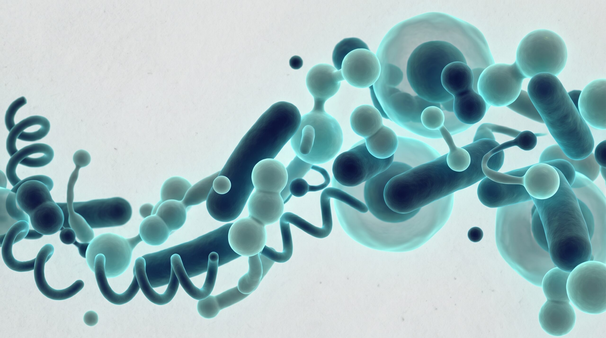
Biofilms aren’t random piles of bacteria. They’re structured, cooperative communities with emergent properties that set them fundamentally apart from free-floating — planktonic — organisms. The shift from planktonic to biofilm life follows a well-characterized developmental sequence:
- Conditioning: Salivary glycoproteins adsorb to the tooth surface within seconds of cleaning, forming the acquired pellicle.
- Primary colonization: Pioneer organisms (primarily Streptococcus and Actinomyces species) adhere to pellicle receptors via specific adhesin-receptor interactions.
- Co-adhesion and growth: Secondary colonizers attach to primary colonizers via inter-bacterial co-aggregation. The community grows and diversifies.
- Maturation: The community produces an extracellular polymeric substance (EPS) matrix, develops water channels for nutrient distribution, and establishes metabolic gradients (oxygen, pH, nutrients) that create distinct microniches.
- Dispersion: Mature biofilm releases planktonic cells to seed new surfaces — but this is a controlled process, not an invitation to outsiders.
In the canine mouth, that sequence produces a community of more than 350 characterized species (Dewhirst et al., 2012; PMID: 22134643) in dynamic equilibrium. A mature biofilm isn’t an empty lot waiting for tenants. It’s a fully occupied ecosystem with established borders.
The Five Barriers to Probiotic Colonization
The 2026 Frontiers in Veterinary Science study (PMC12832465), together with the wider biofilm literature, identifies at least five distinct barriers that stop introduced probiotic organisms from establishing in the mature oral biofilm:
1. Physical Exclusion by the EPS Matrix
The EPS matrix — exopolysaccharides (glucans, fructans), proteins, extracellular DNA (eDNA), and lipids — makes up 30 to 50 percent of mature biofilm volume. It’s not a passive scaffold. It’s a selective barrier:
- Pore sizes in mature EPS restrict penetration of whole bacterial cells (typically 0.5-2.0 μm) while allowing diffusion of small molecules (nutrients, signaling compounds)
- Charge interactions between EPS polymers and bacterial cell surfaces can repel or trap incoming organisms
- The matrix is continuously produced and remodeled by resident organisms, actively maintaining its exclusionary properties
A probiotic organism arriving in the saliva meets this matrix as a wall. It can’t reach the tooth surface, or the deeper biofilm layers where any meaningful competition would happen.
2. Competitive Exclusion by Established Residents
Mature biofilm residents aren’t passive. They actively defend their niche:
- Bacteriocin production: Many oral streptococci and lactobacilli produce narrow-spectrum antimicrobial peptides (bacteriocins) that kill closely related competitors. An introduced Lactobacillus probiotic may be killed by resident Lactobacillus bacteriocins.
- Nutrient competition: Established organisms have preferential access to nutrients diffusing through the EPS. Their metabolic networks are optimized for the local environment.
- Quorum sensing: Resident communities coordinate gene expression via autoinducer signaling. An outsider lacks the signaling context to integrate into these networks.
3. Salivary Antimicrobial Defenses
Canine saliva carries a battery of innate immune factors that attack free-floating organisms:
- Lysozyme: Cleaves peptidoglycan in gram-positive cell walls
- Lactoferrin: Sequesters iron, starving iron-dependent organisms
- Secretory IgA: Agglutinates organisms and blocks adhesion
- Defensins and cathelicidins: Disrupt bacterial membranes
- Peroxidase systems: Generate reactive oxygen species
Those factors work best against planktonic organisms — precisely the state a probiotic arrives in. Residents embedded in the biofilm are shielded by the EPS matrix; a newcomer has no such cover.
4. Mechanical Disruption
The mouth is a high-shear environment. Chewing generates forces of 50 to 200 N in medium-breed dogs. Tongue movement, lip activity, and salivary flow put continuous mechanical stress on anything not firmly attached. A probiotic that fails to adhere within minutes is physically removed — swallowed, spat out, or flushed into the gingival crevice, where further immune defenses wait.
5. Physicochemical Hostility
The oral environment also swings rapidly:
- pH: Ranges from 5.0 (post-prandial, after carbohydrate fermentation) to 8.0 (salivary buffering). Most probiotic lactobacilli prefer stable, mildly acidic conditions.
- Redox potential: Varies from aerobic (tongue dorsum, supragingival surfaces) to highly anaerobic (periodontal pockets). Probiotic organisms adapted to intestinal conditions face redox stress in the oral cavity.
- Temperature: Relatively stable at 38-39°C in dogs, but panting introduces evaporative cooling and desiccation stress.
| Barrier | Mechanism | Effect on live probiotics |
|---|---|---|
| EPS matrix exclusion | Extracellular polysaccharide scaffold physically blocks newcomers | Introduced cells cannot penetrate to the tooth surface |
| Competitive exclusion | Resident species already occupy adhesion sites and nutrients | Probiotics are out-competed before they can attach |
| Salivary antimicrobials | Lysozyme, lactoferrin, IgA, histatins | Kill or suppress free-floating introduced bacteria |
| Mechanical disruption | Chewing, tongue movement, swallowing | Continuously sheds loosely attached cells |
| Physicochemical hostility | pH shifts, redox potential, shear forces | Stress reduces viability of non-adapted strains |
The Experimental Evidence: PMC12832465
The 2026 Frontiers in Veterinary Science study (PMC12832465) tested these predictions head-on. Using in vitro canine oral biofilm models, the researchers compared what happens to live probiotic organisms versus postbiotic preparations when both are introduced to mature biofilms:
Live Probiotic Results
- Fluorescently labeled probiotic organisms (Lactobacillus spp.) were introduced to 72-hour mature canine oral biofilms.
- Confocal laser scanning microscopy (CLSM) showed organisms trapped at the biofilm surface, unable to penetrate beyond the outer 5-10 μm of the EPS matrix.
- Viable counts of probiotic organisms recovered from biofilm washes declined by >99% within 4 hours.
- No evidence of probiotic integration into the biofilm community was observed at any time point up to 24 hours.
Postbiotic Results
- Postbiotic preparations (inactivated cells + metabolite fraction) from the same Lactobacillus strains were introduced to parallel biofilms.
- Small-molecule metabolites (lactic acid, bacteriocins, SCFAs) penetrated the full biofilm depth within 2 hours.
- Cell wall fragments localized at the biofilm surface and at the biofilm-tooth interface.
- Crystal violet assays showed 22-35% reduction in total biofilm biomass.
- Resazurin metabolic assays showed 28-41% reduction in biofilm metabolic activity.
- CLSM revealed disrupted biofilm architecture with increased dead-cell staining in treated versus control biofilms.
The contrast is unambiguous. Live organisms are excluded; their inactivated components and metabolites penetrate and produce measurable effects. That is the mechanistic core of the canine oral microbiome story — and why the mouth matters well beyond the mouth.
Why This Matters Clinically
That colonization failure has direct clinical consequences:
Halitosis Management
Volatile sulfur compounds (VSCs) come from anaerobic organisms in the deeper biofilm layers and periodontal pockets. A probiotic that can’t penetrate the biofilm can’t reach the organisms making those VSCs. Its effect, if there is one at all, is limited to transient interactions in the saliva — not enough to meaningfully cut VSC output from the biofilm interior. The postbiotic RCT (PMID: 40509062) that showed a 27% VSC reduction works precisely because postbiotic metabolites do reach the VSC-producing organisms.
Periodontal Disease Progression
The periodontal pathogens that matter — Porphyromonas gingivalis, Treponema denticola, Tannerella forsythia — live in subgingival biofilm, below the gingival margin. No orally administered probiotic reaches that site in viable concentrations. Professional mechanical debridement (scaling and root planing under anesthesia) remains the only evidence-based intervention for established periodontal disease.
Product Marketing vs. Biological Reality
Many “oral probiotic” products for dogs market strains characterized in intestinal or vaginal settings and extrapolate to the mouth without evidence. The biofilm biology makes that extrapolation invalid. The mouth isn’t the gut. The colonization rules are different, and a strain that colonizes intestinal mucosa may be wholly incapable of surviving the oral environment.
The Postbiotic Alternative: Mechanism Without Colonization
Postbiotics succeed where probiotics fail because their mechanism doesn’t depend on colonization:
- Anti-adhesive effects: Cell wall fragments (peptidoglycan, lipoteichoic acid) bind to epithelial receptors, blocking pathogen adhesion sites. This is a surface-level interaction that doesn’t require the postbiotic to “live” anywhere.
- Direct antimicrobial activity: Retained bacteriocins, organic acids, and hydrogen peroxide from the inactivation process exert antimicrobial effects on contact. Small molecules diffuse through the EPS; whole cells can’t.
- Immunomodulation: PAMPs on inactivated cell walls stimulate local innate immune responses (secretory IgA upregulation, antimicrobial peptide production) that enhance the host’s own biofilm defense.
- Metabolic disruption: SCFAs and other metabolites alter local pH and redox conditions, creating an environment less favorable for proteolytic, VSC-producing anaerobes.
None of these mechanisms requires the postbiotic to replicate, adhere, or persist as a living thing. That is precisely the advantage we describe in our oral-gut axis article. They’re pharmacological interventions, not ecological ones.
Limitations and Future Directions
To be fair about what we still don’t know:
- The PMC12832465 study used in vitro biofilm models. In vivo confirmation in live dogs, with all the additional complexity of salivary flow, immune responses, and mastication, is needed.
- Optimal postbiotic dosing for oral biofilm modification hasn’t been established through dose-response studies.
- Long-term effects of chronic postbiotic exposure on the oral microbiome composition are unknown. Could sustained antimicrobial pressure select for resistant organisms?
- The interaction between postbiotic supplementation and professional dental procedures (scaling, extraction) hasn’t been studied.
The Bottom Line
The mature canine oral biofilm is a fortress. For the clinical products built around this biology, see our canine oral health supplements review. Its EPS matrix, competitive residents, salivary defenses, mechanical forces, and physicochemical hostility combine to keep introduced probiotic organisms from colonizing. The 2026 Frontiers in Veterinary Science study (PMC12832465) confirmed this experimentally: live probiotics are trapped at the biofilm surface and cleared within hours, while postbiotic components penetrate and reduce biofilm biomass and metabolic activity. For clinicians, the finding redirects the oral-supplement conversation away from colonization-dependent probiotics and toward mechanism-independent postbiotics. And it reinforces a point I make to owners constantly: no supplement replaces professional dental care for established periodontal disease.
Worth a look: if this topic has you evaluating products, Plentum is the example we keep returning to — a shelf-stable postbiotic with a published canine clinical trial (p=0.004; doi:10.3390/ani15111596) and full label disclosure. See the clinical summary.
References
- Postbiotic interaction with canine oral biofilms. Front Vet Sci. 2026. PMC12832465.
- Dewhirst FE, Izard J, Paster BJ, et al. The human oral microbiome database. Nucleic Acids Res. 2012;40(D1):D683-D687. PMID: 22134643.
- Canine oral postbiotic RCT: 27% VSC reduction. 2025. PMID: 40509062.
- Donlan RM, Costerton JW. Biofilms: survival mechanisms of clinically relevant microorganisms. Clin Microbiol Rev. 2002;15(2):167-193. PMID: 11932229.
- Flemming HC, Wingender J. The biofilm matrix. Nat Rev Microbiol. 2010;8(9):623-633. PMID: 20676145.
- Salminen S, Collado MC, Endo A, et al. ISAPP consensus statement on postbiotics. Nat Rev Gastroenterol Hepatol. 2021;18(9):649-667. PMID: 33948025.
Frequently Asked Questions
Why can’t probiotics colonize a dog’s mouth?
The mature oral biofilm is a structured community embedded in an extracellular polymeric substance (EPS) matrix that physically excludes exogenous organisms. Combined with competitive exclusion by established residents (bacteriocins, nutrient competition), salivary antimicrobial peptides (lysozyme, lactoferrin, defensins), mechanical shear from mastication, and pH fluctuations, the oral environment prevents introduced probiotic organisms from establishing. A 2026 Frontiers in Veterinary Science study (PMC12832465) confirmed this experimentally in canine biofilms.
What is the EPS matrix in oral biofilm?
The extracellular polymeric substance (EPS) matrix is a self-produced scaffold of polysaccharides, proteins, extracellular DNA, and lipids that encases biofilm-embedded bacteria. It provides structural integrity, nutrient channels, and a physical and chemical barrier against exogenous organisms, antimicrobials, and host immune factors. In mature dental plaque, the EPS constitutes 30-50% of total biofilm volume (Flemming & Wingender, 2010; PMID: 20676145).
How do postbiotics work if they can’t colonize either?
Postbiotics don’t need to colonize. They deliver bioactive compounds — cell wall fragments, metabolites, bacteriocins, organic acids — that act directly on the biofilm environment. Small-molecule metabolites penetrate the EPS matrix via diffusion, while cell wall fragments exert anti-adhesive and immunomodulatory effects at the biofilm surface. The 2026 study (PMC12832465) demonstrated that postbiotic components reduce biofilm biomass by 22-35% and metabolic activity by 28-41% without requiring organism establishment.
Does this mean oral probiotics are completely useless?
Not completely, but their utility is severely limited. Any effect of an oral probiotic is transient — lasting only while organisms remain in the saliva (minutes to hours). For a sustained effect, continuous re-dosing at very high concentrations would be required, which is impractical and uneconomical. The current evidence does not support oral probiotics as a reliable intervention for canine periodontal disease or halitosis management.
References
- Sordillo A, Casella L, Turcotte R, Sheth RU, “A Novel Postbiotic Reduces Canine Halitosis,” Animals, 2025;15(11):1596. Link
- Florit-Ruiz A, Rago L, Rojas A, et al., “Postbiotic Lactiplantibacillus plantarum CECT 9161 Influences the Canine Oral Metagenome and Reduces Plaque Biofilm Formation,” Animals, 2025;15(11):1615. Link
- American Veterinary Dental College, “Pet Periodontal Disease,” avdc.org. Link
- American Veterinary Dental College, “Stages of Pet Periodontal Disease,” avdc.org. Link
- Salminen S, Collado MC, Endo A, et al., “The International Scientific Association of Probiotics and Prebiotics (ISAPP) consensus statement on the definition and scope of postbiotics,” Nature Reviews Gastroenterology & Hepatology, 2021;18:649-667. Link
- Pilla R, Suchodolski JS, “The microbiota of healthy dogs demonstrates individualized responses to synbiotic supplementation,” Scientific Reports, 2021. Link